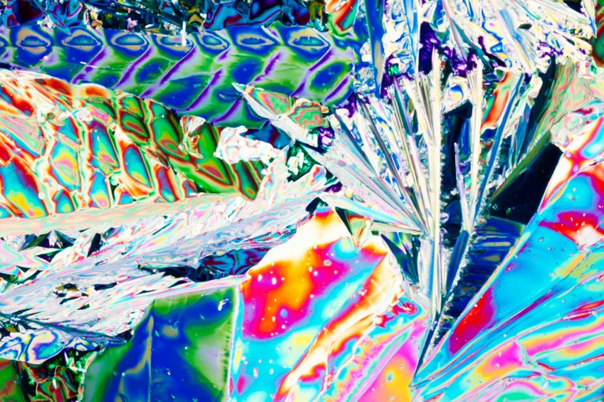
“Beauty in things exists in the mind which contemplates them.” — David Hume.
Scientists who use microscopy are often fascinated by the amazing beauty and complexity of the world on the micro scale. But since they are typically focusing on the data provided by these images, they forget the beauty. Turning microscopy into art is sometimes one step away. Here is how BevShots tell the story:
In 1992, a research scientist named Michael Davidson stumbled upon a genius idea right under his nose — literally. In his 25 year career through the many facets of microscopy, he had taken photographs under the microscope of a collection of items – DNA, biochemicals and vitamins. Looking for novel ways to fund his Florida State University lab, Davidson decided to take his microphotographs to businesses for possible commercial opportunities. While presenting his pictures to established retail companies, one necktie manufacturer changed his creative direction with just one word — cocktails. With this new direction, Davidson took his microphotography a step further […]
Polarized microscopic images of cocktails, brews, and wines formed the Molecular ExpressionsTM Cocktail Collection of BevShots. The images are presented as art prints, neckties, scarves, and beach sarongs, and on glasses, coasters, and hip flasks. Not only they are all breathtakingly beautiful, with amazing variation of colors, they are scientifically accurate, revealing the molecular structure of a crystallized drink.
How are these images obtained? With polarized microscopy, which belongs to the optical microscopy family. The process uses polarized light to illuminate a sample (polarization is the property of an electromagnetic wave, such as light, to oscillate in a certain orientation). A variety of natural and synthetic materials, including polymers, can polarize light. The polarization can be linear, circular or elliptical, in either a clockwise or counterclockwise direction, depending in the properties of the polarizer. Most people are familiar with polarizing materials used in sunglasses, photography filters, and LCD displays. Polarized light is often used in material birefringence analysis for stress analysis.
Polarized light microscopy allows you to see the anisotropic character of a sample. The material is anisotropic if it has different physical properties in different directions. A polarized light microscope has a polarizer positioned before the light reaches the sample, and an analyzer, which is technically a second polarizer, between the sample and the observer (or the camera). The resulting image is obtained through the interaction of polarized light source and an anisotropic sample, in this case, crystalized beverages. Since most of the beverages are multicomponent mixes, the components tend to separate during crystallization, resulting in anisotropic films that are seen as fantastic, multicolor images.
Polarized light microscopy is used to analyze natural and synthetic materials, such as minerals, composites, fibers, ceramics, polymers, polymer films, wood, biological polymers, and tissue samples, and is a popular method in geology, material science, life sciences, and medicine. Turning it into art is certainly a brilliant idea, which might not only appeal to our senses, but also popularize science and share the excitement and awe with everyone.
Image by pilens/123RF.
Source: BevShots, bevshots.com.
Source: “Introduction to Polarized Light Microscopy,” Nikon, microscopyu.com.
Video: About BevShots, by BevShotsMedia, YouTube.com
