“There are things known and there are things unknown, and in between are the doors of perception.” — Aldous Huxley

Our perception of the world is defined to a large extent by what we can see. The human eye has a resolution in the order of 100 micrometers, which is about the thickness of a hair. Using microscopy, whole worlds become visible, inspiring us with beauty and complexity of living, nano, or molecular structures, and providing useful information about the finest details of natural and man-made materials.
Microscopes have been around for a while. The first microscope was built in 1674 in Holland by Anton van Leeuwenhoek, who was the first to observe bacteria and yeast, and the circulation of blood in capillaries. In the 17th century the magnification of microscopes was up to 270x. Toward the end of the 19th century, Ernst Abbe of Zeiss Optical Works calculated the maximum possible resolution for light microscopes.
The limitation of the optical microscopy (OM) comes from the wavelength of light, which is 0.55 micrometers on average. Even using perfect lenses and perfect illumination, light microscopy cannot be used to distinguish objects smaller than half the wavelength of light, or 0.275 micrometers. Present-day instruments have magnifications of up to 1,250x with ordinary light, and up to 5,000x with blue light. To see beyond that, we must illuminate the sample with a shorter wavelength. Using a beam of electrons instead of light allows for higher magnification.
The electron microscope was invented in 1931 in Germany by Max Knoll and Ernst Ruska. To create an image, a beam of fast-moving electrons in a vacuum was focused on a sample, which absorbed or scattered them. The resulting image was captured on an electron-sensitive photographic plate. Because the wavelength of the electrons was significantly smaller than that of photons, the resolution improved dramatically, down to nanometers.
Achievements in microscopy shaped the state of science in the 20th century. Four Nobel Prizes in Chemistry and Physics have been awarded for achievements in microscopy: in 1925 to Richard Zsigmondy, for light microscopy of colloidal solutions; in 1953 to Frits Zernike, for the invention of the phase contrast microscope; and in 1986 to Ernst Ruska, for the design of the first electron microscope, and to Gerd Binnig and Heinrich Rohrer, for the design of the scanning tunneling microscope.
Modern electron microscopy methods are indispensable for biology, medicine, material science, and nanotechnology. They include transmission electron microscopy (TEM) and scanning electron microscopy (SEM). TEM, which is used to study ultra-thin sections of 50-60 nanometers, allows scientists to observe and analyze internal microstructures with magnification of up to 1,500,000x, or 800 times greater than the best light microscope.
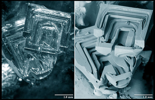
Contrary to TEM, SEM is used to study the surfaces of solid objects, which are scanned by a fine beam of electrons. SEM allows greater depth of focus than the optical microscope and can produce an image that is a good representation of a three-dimensional sample. The images above compare optical microscopy and SEM, with the later providing much higher resolution, even when compared on the same level of magnification.
More recently a different type of sample illumination has been developed by scientists from MIT and NASA, this time using a focused beam of neutrons rather than a beam of light or electrons. The ability to focus the neutron beam using was demonstrated by Boris Khaykovich and co-authors in 2011. The process uses axisymmetric focusing mirrors — consisting of a confocal ellipsoid and hyperboloid, known as a “Wolter mirror configuration,” which is commonly used in x-ray telescopes. Two years later, the small-angle neutron scattering (SANS) prototype was built. After publication in Nature Communications, the new kind of the microscope was described in MIT News last autumn:
Among other features, neutron-based instruments have the ability to probe inside metal objects — such as fuel cells, batteries, and engines, even when in use — to learn details of their internal structure. Neutron instruments are also uniquely sensitive to magnetic properties and to lighter elements that are important in biological materials.
As there is no limit to human desire to see through matter, there appears to be no limit to the technological means to satisfy this curiosity. Science is driven by it.
Source: History of Microscopes. A timeline covering the history of microscopes, by Mary Bellis, inventors.about.com
Source: Microscopes: Timeline, nobelprize.org
Source: Demonstration of a novel focusing small-angle neutron scattering instrument equipped with axisymmetric mirrors, Dazhi Liuet al., Nature Communications, Volume:4, Article number:2556 ; DOI:doi:10.1038/ncomms3556, 30 September 2013
Source: New kind of microscope uses neutrons, David L. Chandler, MIT News Office, web.mit.edu, October 3, 2013
Source: From x-ray telescopes to neutron scattering: using axisymmetric mirrors to focus a neutron beam, B. Khaykovich, et al., Nucl. Instr. and Meth. A 631, 98 (2011); doi:10.1016/j.nima.2010.11.110
Source: Spiegelsysteme streifenden Einfalls als abbildende Optiken fur Rontgenstrahlen, H Wolter, Ann. Der Physik. 10 (1952) 52
Image: Comparison of depth hoar crystal using light (video) and Low Temperature Scanning Electron Microscopy, Wikimedia Commons, Public Domain
