
Electron Microscopes Empower Wide Range of Research
Since the first prototype electron microscope was constructed by German scientists Ernst Ruska and Maximillion Knoll in 1931, electron microscopy has been expanding our understanding of the world on the nano scale. Unlike optical microscopes, electron microscopes use a beam of electrons instead of a beam of light to obtain an image. Electrons are produced by an electron gun, typically a tungsten source (also used in the cathode tubes of older TVs and computer monitors), accelerated and focused by electrostatic and electromagnetic lenses. Solenoids are used to conduct electrical current. The faster the electrons (the lower the frequency), the higher the resolution.
What happens to the electron beam next depends on the type of electron microscopy used. In transmission electron microscopy, or TEM, the electrons go through a very thin (60-90nm) sample. In scanning electron microscopy, or SEM, the electron beam causes the emission of secondary electrons from the surface of the materials, which compose the resulting image. In scanning transmission electron microscopy (STEM), a combination of the two methods, the thin sample is scanned through with the electrons, while in reflection electron microscopy (REM), a reflected beam of scattered electrons is registered. Thin samples that are partially transparent to the electrons are analyzed by TEM or STEM, while surface analysis is best performed by SEM and REM.
Nano Scale
The image obtained by an electron microscope is magnified up to 10 million times, allowing us to see nanoparticles, fibers, cells, components of a cell, or even a molecule of DNA. We can see the faces of tiny creatures like ants and dust mites, and analyze the dust on butterfly wings, as in the video below:
Electron microscopy empowers many areas of science, technology, and medicine. It is indispensable for modern materials science, and is used to analyze a wide range of natural and industrial materials. Electron microscopy can even be used for meteorite analysis, allowing the detection of unique deformation microstructures resulting from collisions that took place millions of years ago.
Electron Microscopy Advances
Recent advances in electron microscopy have started to overcome an inherent drawback, the need for a sample to be dry, solid, immobile — and for most methods — in a vacuum. Conventional sample preparation has depended on the method, and might involve dehydration, fixation, embedding, sectioning, or freeze-fracture. Now, the new development, in situ molecular microscopy, allows the visualization of biological complexes in a liquid environment.
In an article in Lab on a Chip, scientists from Virginia Tech describe a novel approach to visualizing viral assembly in liquid using TEM, with a microfluidic chamber that is isolated from the vacuum system:
We present a novel microfluidic platform to examine biological assemblies at high-resolution. We have engineered a functionalized chamber that serves as a “nanoscale biosphere” to capture and maintain rotavirus double-layered particles (DLPs) in a liquid environment. The chamber can be inserted into the column of a transmission electron microscope while being completely isolated from the vacuum system. This configuration allowed us to determine the structure of biological complexes at nanometer-resolution within a self-contained vessel. Images of DLPs were used to calculate the first 3D view of macromolecules in solution. We refer to this new fluidic visualization technology as in situ molecular microscopy.
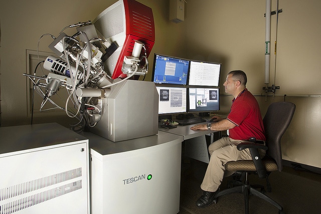
Image by Idaho National Laboratory.
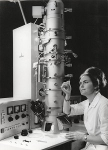
Image by Tekniska museet.
Source: “What Is Electron Microscopy?” by John Innes Centre, jic.ac.uk.
Source: Video, “THIS IS A BUTTERFLY! (Scanning Electron Microscope)” by SmarterEveryDay, youtube.
Source: “Applications of Electron Microscopy in Medicine,” imaging-git.com.
Source: “Messengers From Space, a Scanning Electron Microscopy Investigation,” imaging-git.com, October 10, 2013.
Source: “In Situ Molecular Microscopy. Imaging Viruses in Liquid at the Nanoscale,” imaging-git.com, July 31, 2013.
Source: “Visualizing Viral Assemblies in a Nanoscale Biosphere,” by B.L. Gilmore et al., Lab on a Chip, RCS Publishing, 13: 216 (2013), DOI: 10.1039/c2lc41008g.
